Are you tired of the constant pain and discomfort caused by bunions? Do they make even the simplest activities (like walking or wearing shoes) feel impossible? Don’t let bunions dictate your life.
At Gulf South Foot & Ankle, we believe you shouldn’t have to compromise your lifestyle because of bunion pain. Our expert podiatrists provide both conservative and surgical solutions to help you move freely again.
Book an Appointment today
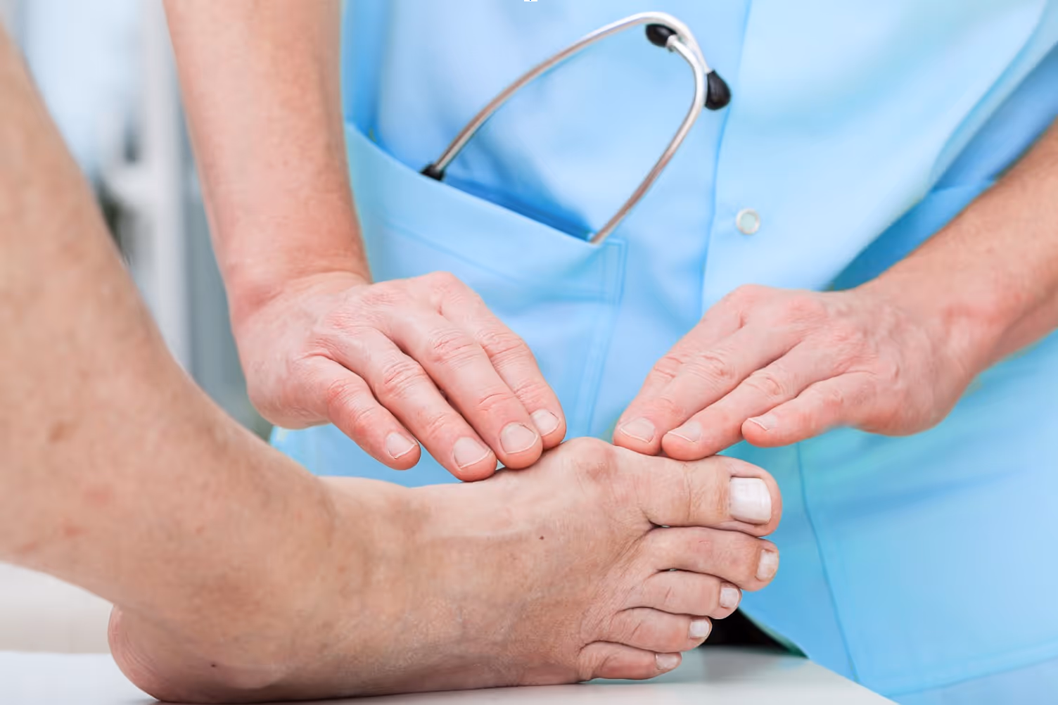
A bunion forms when the big toe pushes against the next toe, forcing the joint out of alignment. Over time, the joint sticks out, becomes irritated, and can lead to long-lasting pain and stiffness.
Bunions are not just a cosmetic issue, they are a progressive foot deformity that can:
At Gulf South Foot & Ankle, we don’t just mask symptoms, we address the root cause of bunion pain so you can stay active without constant discomfort.
Contact us now for helpRecognizing bunion symptoms early can help you prevent progression. Common signs include:
If you notice these symptoms, seeking professional bunion treatment is the best way to protect your foot health.
Ignoring bunions won’t make them go away, in fact, they often get worse with time. Untreated bunions can:
Early treatment helps prevent long-term complications and may reduce the need for surgery.
Every foot is unique, and so is every bunion. That’s why we tailor every treatment plan to your needs and lifestyle. Your care may include:
When conservative methods are no longer effective, our specialists offer minimally invasive bunion surgery. Surgery can:
Our surgeons use advanced techniques designed to shorten recovery times and maximize long-term results.
Healing from treatment depends on the severity of the deformity and the treatment chosen. Many patients who undergo non-surgical bunion care notice relief within weeks. Surgical patients typically experience improved comfort, shoe fit, and mobility once recovery is complete.
Our goal is always the same: to help you return to the activities you love like walking, exercising, or simply living without bunion pain.
Living with bunion pain doesn’t have to be your reality. Whether you need bunion treatment or are considering bunion surgery, our team is ready to help you take back your comfort and confidence.
Book an Appointment Today